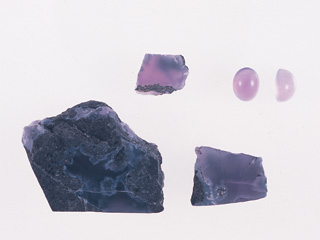
We recently had an opportunity to identify a violet translucent stone on the photo 1. The stone was, after our investigation, proved to be a new variety, natural colour chalcedony containing iron. The background and gemmological features of the stone are reported here.
Introduction
Chalcedony is cryptocrystalline quartz constructed by minute crystals of ¿(alpha)-quartz, and it has many colour varieties such as white chalcedony, red chalcedony (carnelian) or green chalcedony (chrysoprase and mtorolite). A violet variety is quite rare and most of natural colour materials are actually violet-blue. A variety called amethystine chalcedony is violet, but its transparency and saturation of the colour are commonly low.
The name chalcedony comes from Chalceton, a town in Greece from where the material occurred. Its Japanese name is gGyoku-zuih and those with colours arranged in stripes or concentric pattern are called gMenouh (agate). Chalcedony, as well as agate, occurs in each part of the world, and its major localities are; Brazil, Australia, Madagascar, India and Poland. Domestically, it is produced in coastal area of Hokkaido, Fukui, Ishikawa and Chichijima islands. It commonly forms in shapes such as mass (botryoidal), vein or sphere to hemisphere, and it occurs in cavities in igneous rocks (basalt and dolerite) as well as dykes of metamorphic rock or granite pegmatite and hydrothermal vein. According to Mr. Minoru Kameyama of MIYUKI CO., LTD., who presented the sample stone, this violet chalcedony is a new product from Indonesia in East-South Asia, although its detailed locality is remain unknown.
Gemmological Features
The sample used in this study is shown in the Photo 1. Minute netted chalcedony crystals are filling inside of a cavity of a black host rock. They are commonly composed of gbotryoidalh groups of minute quartz and their deep violet colour remind you of amethyst or violet sapphire. A part showing beautiful colour and transparency is cut cabochon for gem use.
Gemmological properties of the violet chalcedony are listed in the Table 1.@
 |
|
Features |
| Colour |
Violet |
| Transparency |
Translucent
|
| Hardness |
V |
| RI |
1.540|1.543 |
| DR |
0|0.003 |
| SG |
2.62 |
| LWUV |
Blueish white fluorescence
|
| SWUV |
Inert
|
| Colour filter |
Dark red
|
|
| Photo 1: |
A violet chalcedony from Indonesia |
|
| Table 1: |
Gemmological properties of a violet chalcedony from Indonesia |
|
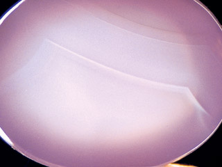
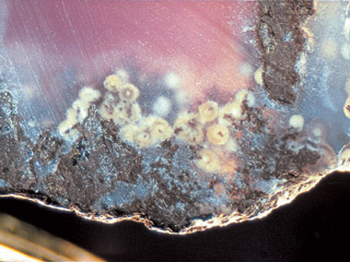
Its RI was 1.540-1.543 with a small DR (0.003) and its SG was 2.62. No pleochroism was observed as it was polycrystalline, and it appeared red through a colour filter. It showed blueish white fluorescence under LWUV, while it was inert to SWUV. No characteristic absorption was observed under a handy-type spectroscope. As shown in the Photo 2, distinct colour patches of agate-type (which is never observed in amethystine chalcedony) were observed under magnification. Characteristic strain was not observed under crossed polarised light while immersing the sample in water. White spherical crystal aggregate was observed around the boundary between the chalcedony and its host rock (Photo 3), and Raman spectroscope detected a peak on 463cm-1 due to Si-O-Si molecular vibration. X-ray fluorescence analysis on minute areas revealed that the main constituent of the material was SiO2 and impurities such as Fe, Ca, Mg and Mn were included. Considering the nature of the host rock that is going to be described later, this white spherical crystal is presumably cristobalite.
 |
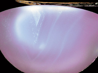 |
| Photo 2: Agate-type growth colour zoning distributed in the chalcedony |
 |
 |
| Photo 3:White spherical crystal aggregate ? cristobalite |
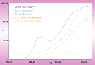
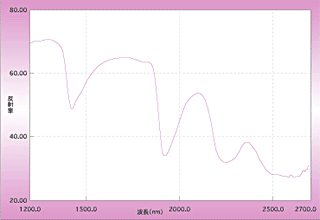
Spectral analysis in visible-UV region showed a broad absorption around 540 nm from yellow to green area on a spectrum probably due to iron and a small absorption around 360 nm in UV region (Fig. 1). The region between blue and violet is a transmission area, which makes the colour of the stone characteristic. Contrary to this, blue chalcedony shows an absorption around 590 nm, and pale brown, pale green and pale white chalcedonies only show simple decline of transmittance from infrared to UV region. Near infrared (NIR) spectral analysis gave us a spectral pattern typical to chalcedony (Fig. 2), in which a vibration absorption on 2254 nm due to Si-OH, a vibration absorption on 1924 nm due to molecular water (H2O) and an absorption on 1426 nm due to hydroxyl (OH) were detected.
 |
 |
| Figure 1: |
Transmission spectra of chalcedonies in each colour in UV-visible region |
|
| Figure 2: |
Reflectance spectra of violet chalcedony in near infrared region |
|
|


