|
|||||||||||||||||
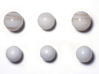
| ABSTRACT Pearl nuclei made of freshwater bivalve Anodonta Woodiana and saltwater bivalve Tridacna Squamosa were analysed by LA-ICP-MS analytical method and the results were considered for the material identification of nuclei of those shells used in cultured Akoya pearls. The distinction between nuclei themselves of these two shell materials can be easily carried out by X-ray fluorescence analysis. However, cultured pearls using nuclei of these material cannot be analysed their nuclei directly by the method because the nuclei are all covered by nacre. LA-ICP-MS can detect each layer of nacre on a pearl from its surface toward a nucleus by irradiating a laser beam continuously and it can eventually analyse the nucleus inside the pearl to identify the material. INTRODUCTION In pearl cultivation, insertion of a piece of mantle into a pearl oyster is inevitable and the piece is said to act a big role to decide body colour and quality of a pearl. As for a nucleated cultured pearl, a nucleus (a spherical bead) inserted with a piece regulates shape of a pearl sac and it also influences the size and shape of the pearl produced. For this reason a nucleus is required to be a sphere and to have property to resist unique fashioning of pearl and aging. Various materials such as glass, marbles or corals have been tried for a nucleus of cultured pearl, but a nucleus of the Anodonta mussel, which is made of the freshwater bivalve Unionidae (Photo-1, top) have been used for a long period because of its good compatibility to a mother oyster and its durability. Although depletion of the resources was once concerned for the mussels and substitute nuclei of giant pacific oyster shell that were made into ceramics or other materials were experimentally produced, none of their properties reached to the level of the Anodonta nuclei. In recent years, nuclei made of shell from the saltwater Tridacna Squamosa (Photo-1, bottom) have been reportedly used for Chinese and Tahitian cultured pearls and sold on the market. The Tridacna nuclei are absurdly inexpensive and its use will possibly be expanded in the future. However, the Tridacna is fraught with various problems such as follows:
ü@ü@Tridacna Squamosa is listed in AppendixIIof Washington Convention, and it requires permission on export. Other than the oyster itself as a living organism, nuclei made of this shell and cultured pearls using such nuclei are also included in the list for commercial trade.
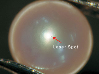
Disadvantageous Properties The most serious problem of the Tridacna nucleus is that it has more propensity than the Anodonta nucleus to crack during drilling process. The crack in a nucleus will consequently result in cracking of a pearl itself. There was a report on pearls with thin nacre that the pearls using the Tridacna nuclei had higher incidence of cracking than the pearls using the Anodonta nuclei (Pearl News No. 145). Definition of a Pearl Nucleus Japan Pearl Promotion Society regulates in its Pearl Standards that pearl nuclei should contain pearl layer with nacreous structure of the same material, density and hardness as the pearl itself (Definition of Pearl - 2-1, Nucleated Cultured Pearl). While the Anodonta has pearl layer with nacreous structure, the Tridacna has a crossed lamellar structure and its density and hardness are not the same as the pearl itself, therefore the use of the Tridacna nuclei goes against the definition above. ü@ü@Distinction between the Anodonta nuclei and the Tridacna nuclei has been made traditionally based mainly on observation of appearance, ultraviolet fluorescence, magnetic susceptibility and magnetic anisotropy. In nucleus identification of a pearl that has been already drilled, for instance, measurement of fluorescency is reportedly possible when ultraviolet fibre optic light of 0.1mm is inserted to a drilled hole (Tanaka, Komatsu; 2004.8 No.153), and for undrilled pearl, a measuring equipment to distinguish these two material with non-destructive method by the difference of magnetism has been developed recently (Camden Corporation 2003.9). Measurement of impurity elements by an X-ray fluorescence analytical device also gives us important clues to distinguish saltwater pearls from freshwater pearls. As freshwater pearls contain more manganese, they can be distinguished from saltwater pearls, likewise the nuclei of these pearls are easily distinguishable. However, cultured pearls using these nuclei grow nacreous layers around themselves and the distinction of the nuclei by their appearance becomes impossible. Elemental analysis by an X-ray fluorescence analytical device cannot be applied to detection of nuclei inside pearls because the analysis is limited only to the surface of a material. ü@ü@The authors have analysed both nuclei material using Laser Ablation-Inductively Coupled Plasma-Mass Spectrometry (LA-ICP-MS) to examine whether their distinction is possible. With the LA-ICP-MS analysis, inspection of a nucleus inside a pearl is possible by irradiating a laser beam continuously to analyse nacre from the surface into depth direction sequentially. A tiny hole of about tens of micrometer in diameter (photo 2) will only be left on the pearl surface after this inspection as a trace of laser irradiation, which is quite different from a laser-drilled hole made on a colourless transparent diamond, and this hardly damage the visual appearance of a pearl. This inspection method is explained below.
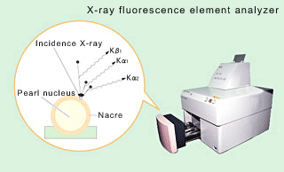
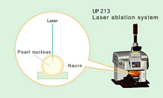
SAMPLE AND METHOD ü@ü@Objects of this study were the Anodonta nuclei, the Tridacna nuclei, Japanese saltwater cultured Akoya pearls containing the Anodonta nuclei, and Chinese saltwater cultured Akoya pearls containing the Tridacna nuclei, and an X-ray fluorescence element analyzer and a LA-ICP-MS device were used for the analysis. The sample of Chinese saltwater cultured Akoya pearls were provided by Mr. Hiroshi KOMATSU of Pearl Science Laboratory. For LA-ICP-MS analysis, a laser ablation system of NEW WAVE RESEARCH was used with laser beam diameter 30 ā╩éŹ,ü@ laser pulse frequency 20 Hz, laser energy 0.107mJ, measurement period 40-120 seconds and number of measurement 3. X-RAY FLUORESCENCE ELEMENT ANALYZER When a substance is irradiated with X-ray, elements consisting the substance emit unique secondary-X-rays. By examining these secondary X-rays, variety and content of each element consisting the substance can be analysed. As a gem mineral consists of unique combination of elements, it can be compositionally analysed non-destructively with this method (Fig 1). For more details, please refer to the GEMMOLOGY March 2003 and July 2003. LASER ABLATION-INDUCTIVELY COUPLED-PLASMA MASS SPECTROMETRY (LA-ICP-MS) LA-ICP-MS is a highly sensitive method to quantitatively and qualitatively analyse constituents of a sample by detecting a mass of grains, which were vaporised and dispersed when the sample was irradiated by a high-energy laser beam of shorter wavelength on its surface, and then ionised by plasma generated by high frequency power (Fig 2). For the details of the instrument, please refer to the GEMMOLOGY November and December 2003 and June 2004, or visit our website http://www.gaaj-zenhokyo.co.jp.
|
|||||||||||||||||
|