|
|||||||||||||||||||
|
The latest topics at the GAAJ laboratory are introduced. This report is based on an oral presentation at the general lecture in the annual meeting of the Gemmological Society of Japan 2006, which was partly added and amended.
Introduction GAAJ laboratory has been operating continuous experiments on HPHT-treated diamond, under the cooperation of Dr. Hisao KANDA of NIMS, and its results were reported in various occasions including the past annual conference of the Gemmological Society of Japan. In the latest operation, we have examined the difference of colour hue, the difference of external and internal features and analysed photoluminescence (described as PL analysis hereafter), before and after the HPHT treatment in two experiment examples. The details are reported. TypeIIb Brown Diamond Colour Hue Alteration TypeIIb brown diamond generally shows pale blue to greyish colour hue, but it may appear brownish if it accepted plastic deformation. The quite unusual typeIIb brown diamond was used this time for HPHT-treatment experiment. Photo 2 shows the appearance before and after the treatment. The stones weighed 0.357ct and 0.389ct and were colour-graded as K and V. L. Brown respectively. They were treated under temperature at 1800ºC and 6GPa pressure for ten to fifteen minutes at NIMS, and subsequently they were treated again at Sundance Diamonds in US on our request (the treatment condition is unclear). The stones weighed 0.349ct and 0.382ct and were colour graded as Light Grayish Blue and Light Blue respectively after re-cutting. The colour alterations were delivered from removal of distortion-induced brown tint by HPHT treatment and blue colouration derived from boron in typeIIb stone.
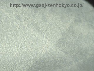
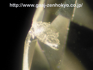
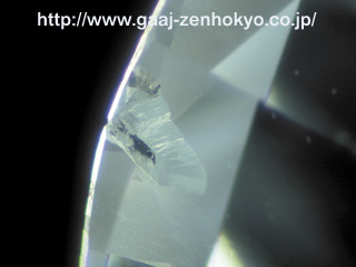
Observational Features After HPHT treatment, diamond surface is fused and gives frosty appearance, so that the stone treated in this process requires re-cut or re-polish afterward. Therefore, when fused mark is recognised on the stone is suspected to HPHT-treated. Contrary to this, when faces such as naturals without this mark are left on a stone, it may highly be untreated. Photo 3 shows a feather that is partly graphitised. Photo 2 shows a round brilliant cut diamond that is broken, which displaying a possibility of damage caused by enlarged cleavage caused by HPHT treatment in lower clarity samples.
PL analysis PL analysis was operated using RANISHAW microscopic Raman spectroscope RAMA-SCOPE SYSTEM 1000B. The 514nm argon laser and 488nm argon laser were used as exciting laser. Natural typeIIb diamond generally shows weak or almost vanished 637nm peak, often with peaks at 566nm and 648nm. Change is mainly seen in N-V centre before and after the treatment, as (negatively charged N-V defect) 637nm peak is likely to become stronger, or peaks at 566nm, 575nm and 630nm existed before the treatment tend to disappear after the treatment. |
|||||||||||||||||||
|


