| Rainbow Garnet The rainbow colour seen in the andradite from this area is assumed to be derived from interference of light produced by thinly layered two substances that have different refractive indices [fig 4] similar to the principle of labradorescence in labradorite. From this theory, the garnet was made into a thin section and its internal structure was investigated. The sample used for this study was a dodecahedral crystal with the size of about 10mm, and it was cut perpendicular to the c-axis (the direction of [001]). The mean chemical composition of the sample was 96% of andradite and 3% of grossular, which shows that the material is very close to the end member andradite.
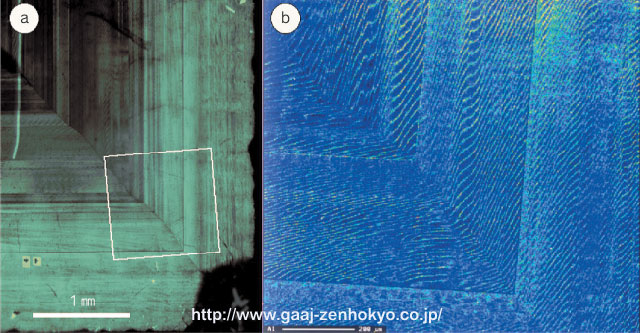
@@ Observation with an optic microscope and an scanning electron microscope revealed that growth bands ran parallel to {110} face and wavy lamellae in 10`20òm intervals developed all over [fig 5]. Difference in composition was seen in this wavy lamellae, and the thicker layers contain less Al (2% of grossular) and several òm thinner layers more Al (8% of grossular) [fig 5b]. Optically, the latter layers seem to have optical anomaly (that is, decrease of symmetry elements from cubic crystal system).
@@ Lamellae structure corresponding to wavy lamellae that form parallel with growth form was seen on the crystal surface [fig 6]. From this observation wave lamellae is assumed a structure that is formed during growth.
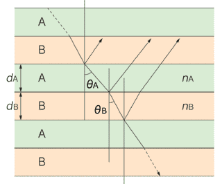
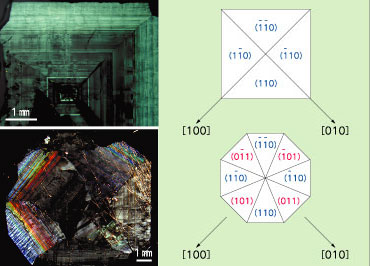
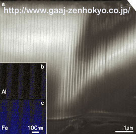
As the wavy lamellae are also seen in rainbow garnet from Adelaide they may be regarded as a structure unique to the rainbow garnet, however, they are not so prominent in rainbow garnet from Sonora. Also size of the structure is much larger than that of wavelength of light, it is unlikely that the structure is the cause of iridescence. @@In the {110} growth sector observed in a thin section that is perpendicular to the c-axis, a sector in which {110} face is intersecting at 45 degree to the plane of the plate (each of (101), (011), (01) and (01) sector in bottom photo in fig 7) shows very strong iridescence. Contrary to this, a sector in which {110} face is intersecting perpendicular to the plane of the thin plate (each of (110), (10), (10) and (0) in fig 7) ? that is, incident light becomes parallel with the {110} face in the sector ? does not show iridescence. From this observation it can be assumed that iridescence is a structural colour caused by multilayer interference that is produced when a light passes through layered thin films parallel to {110} face. This indicates the existence of layered structure that has the interval of about several hundreds nm in thickness, equivalent to the order of the wavelength of light, and that is parallel to {110} face. @@The sample was further analysed with a transmission electron microscope with high resolution power. The result revealed the existence of fine lamellae between wavy lamellae that have quite regular interval and is parallel to {110} [fig 8]. A composition mapping [fig 8b and c] using the HITACHI HD-2300 Scanning Transmission Electron Microscope (courtesy to Naka customer centre, Hitachi Science Systems Ltd.) also showed difference in composition of Al/Fe between the both layers, hence difference of refractive indices between them was indicated. From this result, the cause of iridescence can be concluded as multilayer interference of light produced by these fine lamellae. The fine lamellae continuously across the wavy lamellae that were produced during the growth, so that this structure is assumed to be developed after the growth. As the compositional difference between the layers is distinctive and their interfaces are sharp, the fine lamellae are highly possibly developed by exsolution after the growth of garnet.
Summary The discovery of a large amount of globally rare rainbow garnet in Japan was a big surprise. For some period after the discovery the new material had been known only to mineral fanciers. Once it was on a special exhibition at the 18th Tokyo International Mineral Fair held in Shinjuku in last July and appeared on the Asahi Shimbun on 2 August with a coloured photo, the stone attached national attention. The collection of the stone has been now prohibited but looters do not cease and there was even a person under arrest in this September. The site is inside the National Park and its quiet mountains are listed in the World Heritage Lists. Besides, rainbow garnet in good quality is almost depleted and no gem quality material is left there. We strongly hope people to refrain themselves from illegal digging and be sure not to damage the nature at site. |
||||||||||||||||||||||||||||||
|
||||||||||||||||||||||||||||||