|
||||||||||||||||||||||||||||||||||||||||||||||||||||
| Abstract We reviewed difference on observation between sector zoning of cube-octahedron rarely seen in natural diamond and that commonly seen in synthetic diamond produced by high temperature high pressure method. Under the ultraviolet light test that is used for standard gemstone identification, both natural and synthetic stones show similar sector zoning as a cross-shaped patchy fluorescence. Although those growth sectors and domain of the sectors are difficult to observe under a gemmological microscope, but the difference of sector zoning between natural and synthetic stones can be clearly observed by applying techniques such as X-ray topography or cathodo luminescence. Furthermore, by using an X-ray analytical microscope internal structure of centre-cross diamonds of both natural and synthetic can be observed. Introduction �@�@One of the basic concepts in distinguishing between natural and synthetic diamonds is a difference in morphology. Natural diamonds generally show octahedron crystal form while synthetic diamonds produced by high pressure high temperature method show cube-octahedron. However, natural diamond may occasionally show mixed-habit growth surrounded by cubic faces and octahedral faces (Frank. 1967), and such stone is called a centre cross diamond. During a growth of natural diamond in magma of silica solution, when a growth speed of {100} faces become relatively slower than that of smooth {111} faces, not crystallographically flat {100} faces but curved rough faces coexist with {111} faces and they form a cross-shaped pattern with two different growth sectors (fig 1).
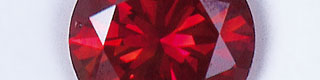
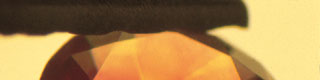
Such `centre-cross' structure is explained due to a shift of supersaturation during the crystal growth (Kitamura 1993). Contrary to this, synthetic diamonds produced by HPHT method grow {111} and {100} faces always as smooth crystal faces in metal solvents such as iron or nickel, resulting the crystal formed in cube-octahedron (Tolansky and Sunagawa 1959). In this research, identification techniques of natural/synthetic diamonds with higher sensitivity are discussed by comparing and observing both natural and synthetic diamonds showing sector zoning to point out similarity and difference between them. Samples and Methods �@�@In this study, one natural diamond showing mixed-habit growth (sample A) and one Chatham-created synthetic diamond (sample B), two pieces in total, were the subject of analysis (Fig 2). They are faceted in brilliant cut and each weighs 0.105ct and 0.213ct with purplish red and orange-red colour. They were analysed by magnification test with a gemmological microscope, UV fluorescence test, cathode luminescence (CL) technique (that is CL attached to a luminoscope and a scanning electronic microscope) and an X-ray analytical microscope. On the observation by a scanning electronic microscope, the samples were coated with gold to avoid their conductivity and their tables were set perpendicular to the electron beam. Accelerating voltage of the electron beam was 20kv. In the analysis by an X-ray analytical microscope, the samples were analysed from the direction almost perpendicular to the table facets without any preparation.
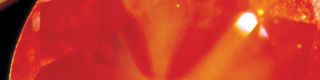
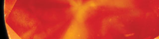
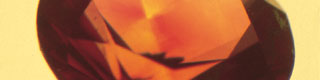
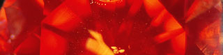
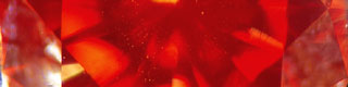
Results and Discussion �@Magnification Test and Observation of UV Fluorescence �@�@As shown in Fig 3a, the sample A contains minute inclusions that are distributed in the centre of the stone, and it shows visible fluorescence of pinkish red colour against the illumination of strong fibre optic light. When observing the stone being immersed in methylene iodide, colour zoning in four directions from the centre of the crystal towards outer edge can be confirmed, and minute inclusions are concentrated and elongated in the crystal direction of {111} in this area (Fig 3b). Pale colour zoning are also observed perpendicular to octahedral faces, as well as colour concentration on a culet that may be due to an irradiation (as the sample is presumed to have been irradiated by electron beam and then annealed). Dark red fluorescence and, especially, cross-shaped patchy fluorescence caused by centre-cross structure (growth sectors) are recognised under long wave ultraviolet light (Fig 4).
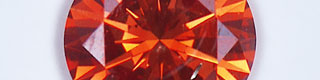
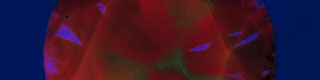
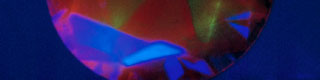
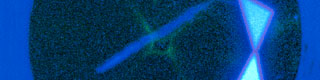
The sample B contains metal inclusions originate in solvents that are characteristic to synthetic diamond, and minute inclusions in the centre of the crystal (Fig 5).As with the sample A, this is also presumed to have been irradiated with electron beam and then annealed. Cross-shaped patchy fluorescence characteristic to synthetic diamond due to sector zoning of cube-octahedron is observed under long and short wave ultraviolet lights (Fig 6).
|
||||||||||||||||||||||||||||||||||||||||||||||||||||
|
||||||||||||||||||||||||||||||||||||||||||||||||||||