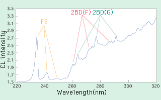
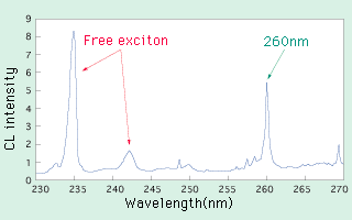
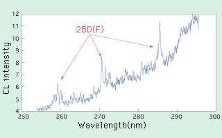
| 3.2 Cathode Luminescence Spectra In the CL spectrum of a typeII natural brown diamond shows groups of luminescence peaks called 2BD(F) and 2BD(G) as shown in fig.-7. They are made of several peaks and lie between 260 and 290nm. Peaks on 260nm, 272nm and 286nm make 2BD(F) and 264nm, 276nm and 290nm peaks make 2BD(G). These groups of luminescence peaks may diminish or disappear when a crystal is HPHT-treated to vanish brown colouration. Particularly, 2BD(G) disappears with low temperature. From this we can expect that the groups of luminescence peaks called 2BD(F) and 2BD(G) were produced by strain similarly to the brown colouration. Several brown crystals have been HPHT-treated and disappearance of 2BD(F) and 2BD(G) was confirmed so far, however, some of them left a sharp peak at 260nm after the groups of peaks disappeared (fig.-8). This raised a question on the 260nm peak that it may also relate to the strain. HPHT treatment has been regarded as a process to remove the strain so far, but it is possible to think that the process produces strain in a crystal because the treatment involves high pressure. Thus we examined on two points listed below by using HPHT-treated stress-free diamonds synthesised with high pressure in the perspective of producing strain: (1) Will the luminescence of 2BD(F) and 2BD(G) appear? (2) Will a peak at 260nm appear? The spectra shown in fig.-9 were obtained by measuring CL of a synthetic crystal A. Those spectra before and after HPHT treatment were shown in (a) and (b). By comparing them, it was studied that the 260nm peak was produced by HPHT treatment. In this test, electron beam was irradiated on a spot as minute as 1 micrometer and each spot of irradiation showed individual spectrum. The 260nm peak was detected in spectra from near edges where have stronger strain in a crystal, and this empirically confirms that the peak relates to strain. Shown in fig.-10 are spectra of a synthetic crystal B before and after HPHT treatment. Jagged numerous peaks are recognised between 260 and 290nm. In comparing to fig.-7, these peaks are read as 2BD(F). They were also produced by HPHT treatment and proved related to strain. Shape of the peaks is more distinct than that in fig.-7, and countless fine peaks are seen in fig.-10. Each wavelength of the fine peaks differs according to the measured spots, and the cause of this group of fine peaks is unknown. In fig 10, no 2BD(G) is seen because it is thought to disappear by the HPHT treatment in this test, as 2BD(G) disappears with rather low temperature. Å@Å@As described above, we were able to confirm empirically that peaks of 260nm and 2BD(F) were produced by creating strain in two crystals with HPHT treatment. From this, we can conclude that 2BD peaks and a 260nm peak seen in natural brown diamond crystals are related to strain. Crystal A and B showed different spectra, but this is thought due to the temperature difference of the treatment. Crystal A must have been treated with higher temperature. More detailed experiment in consideration of temperature value in HPHT treatment should be required hereafter.
4. Conclusion In relating to decolouration of natural brown diamonds by HPHT treatment, changes of cathode luminescence spectra by HPHT treatment were studied. Peaks of 2BD luminescence and 260nm luminescence were empirically confirmed being formed by strain. However, they have different behaviour on appearance / disappearance. A peak of 2BD luminescence is formed in low temperature but disappears in high temperature, whereas a peak of 260nm luminescence is formed in high temperature. |
||||||||||||||||
|


